Doctors at a Sioux Falls hospital have a new tool to help patients facing cancer. Sanford Health now has the world’s smallest microscope. It allows physicians to view cells as they live inside patients.
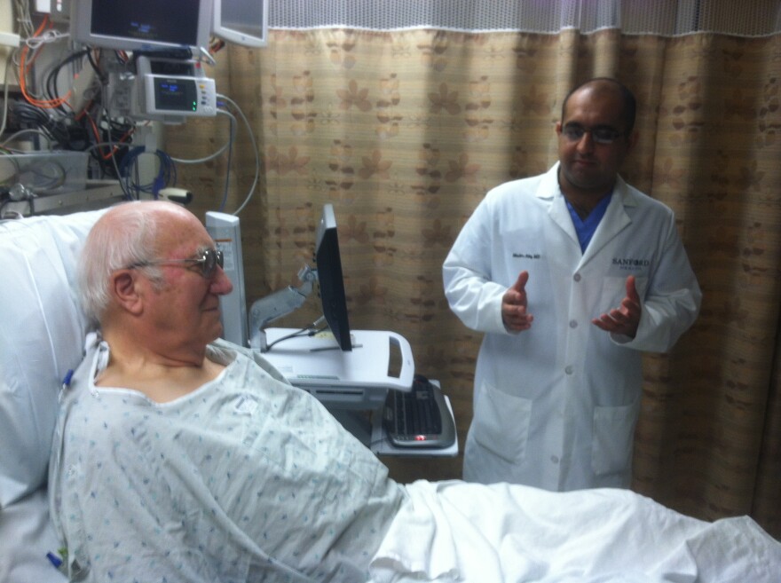
His family huddles along the wall as an aging man stretches out in a hospital bed. They listen to a doctor clad in the classic white coat. A fluorescent light illuminates the room. The patient wears a light patterned hospital gown.
Glen Rabuck from Redfield, SD peers through thick lenses and explains he’s at Sanford Health in Sioux Falls after a referral from his Aberdeen physician.

"I’m here to have a check on cancer in my esophagus," Rabuck says. "I’ve had trouble with acid reflux for 40 years, and now it’s went to Barrett’s, and now they think it’s showed up cancer maybe."
Rabuck is certainly not the only patient checked for cancer, but his case is unique because of the way doctors screen his cells. It happens while the cells still live inside Rabuck’s body.
Once sedated, Rabuck lies on a table in a dimly-lit room. Machines set up around him hum and hiss. Images flash on two large computer monitors. Dr. Muslim Atiq focuses intently on the screens. Blurred images come into focus and show real cells alive in the patient’s esophagus. The doctor notes the images while simultaneously maneuvering a tube fed into the patient’s throat with his right hand and guiding a mechanism with his other hand. That controller operates the world’s smallest microscope.

Atiq is a gastroenterologist, so he works with parts of the body including the esophagus, stomach, small bowel, large bowel, pancreas and liver.
This doctor is one of a handful trained to use the Cellvizio microscope. It’s so tiny that the lens fits on the end of a long tube doctors put inside a person’s body to capture live images of intricate structures.
"It’s like looking at the stars with a telescope. You’re actually on the same principle by looking inside the GI tract with a microscope, just magnifying small things as much as you can," Atiq says.
In this procedure, Atiq searches for abnormal cells in Glen Rabuck’s throat. Atiq explains doctors keep close watch on people diagnosed with a condition called Barrett’s esophagus, because it often leads to cancer.
"Previously we were bringing them back and doing an endoscopy every so many years and then we would target biopsies at that site to get the tissue that we need and the pathologist would look at it for us," Atiq says. "Now we can actually go in, look at an area in real time, and see what areas are abnormal so that we can target our biopsies. And if there is suspicion that the area is abnormal, we can actually make a plan to treat it in the same setting."

Atiq says gathering instantaneous information about abnormal cells has obvious advantages. Doctors can specifically, physically outline malicious material inside the body. The opportunities for accurate diagnoses increase because physicians are less likely to biopsy a healthy area and miss nearby cancer cells. Plus doctors can report immediately that a patient has cancer and begin treatment right away. Sometimes it’s the opposite.
"Somebody comes in and you tell them up front, based on what we see today, we don’t see anything abnormal, and that’s a big relief. Because if we can put ourselves in that same scenario, that’s some big news that you’re telling someone rather than waiting for the next week or so to get an answer," Atiq says.
Atiq says finding a person doesn’t have cancer is often bigger news than confirming suspicion a patient is sick. That’s not Glen Rabuck’s case.
Experts viewing the cells living in his throat confirm Rabuck’s condition has escalated. Doctors identify abnormal cells and still test to confirm the cancer. But the view from the mini microscope lets Doctor Atiq inform the patient and his family right away. Now they can move forward with a plan to tackle the disease.

Rabuck says he isn’t sure how he feels about being one of the first patients to have the world’s smallest microscope searching through his cells.
"I haven’t thought about that! I don’t know. Important, I guess," Rabuck says. "I suppose I could go down in history, eventually. I don’t know."
Rabuck says his exam is valuable, not for only his health but because his test case offers opportunities for other patients.


